Mammograms, Breast Ultrasounds and MRI's: What's It All Mean?
 The key to saving lives from breast cancer is early detection. The American College of Radiology recommends annual mammogram screening beginning at age 40 for women of average risk. Higher-risk women should start screening earlier. The purpose of a mammogram is to assist in detecting breast cancer sooner that in can be felt in the breast.
Mammography
Mammography is considered to be the gold standard and is the best tool for detecting all shapes, sizes and forms of breast cancer. A mammogram uses a dedicated x-ray machine to take images of the breast. The images are evaluated by the Radiologist who will look for subtle changes in the breast tissue, which may be an indicator of developing cancer.
Having routine mammograms is the best tool to detect breast cancer early.
A change in the breast tissue may appear as a small density, or as microcalcifications that show on the x-ray image. Any time these changes are seen on a mammogram, the Radiologist will want to follow with additional images to further evaluate. If you are asked to return after your screening mammogram, it does not necessarily mean you have cancer; it just means that additional information is needed to make an accurate diagnosis.
Breast Ultrasounds
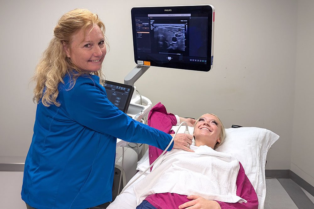
 Breast ultrasounds are considered to be a good complimentary tool that helps to evaluate an abnormality that may be seen on a mammogram. It can help decide if the area in question is a cyst (a fluid filled sack), or a solid mass (which might need further testing to be sure it is not cancer).
A breast ultrasound is a good secondary method of evaluating the breast but is not used by itself. It can't detect the small subtle changes that a mammogram can. Ultrasound is best used when you know the exact area in question, not to scan the entire breast.
Magnetic Resonance Imaging (MRI)
MRI is a great modality to use when diagnosing small abnormalities in the breast. A woman that is at high risk for breast cancer may benefit by having a screening mammogram done followed by an MRI 6 months later and repeating this cycle. The combination of these two tests will help to pick up small changes in the breast tissue.
MRI testing is also helpful when there is known cancer in one of the breasts. The surgeon will want to use an MRI scan to look at the extent of the cancer and if or how much of the other breast is involved. Mammograms and breast ultrasounds aren't always able to pick up the extent of just how invasive the cancer is, so an MRI is used to help the surgeon recommend if a small portion or the entire breast should be removed.
3D Mammography
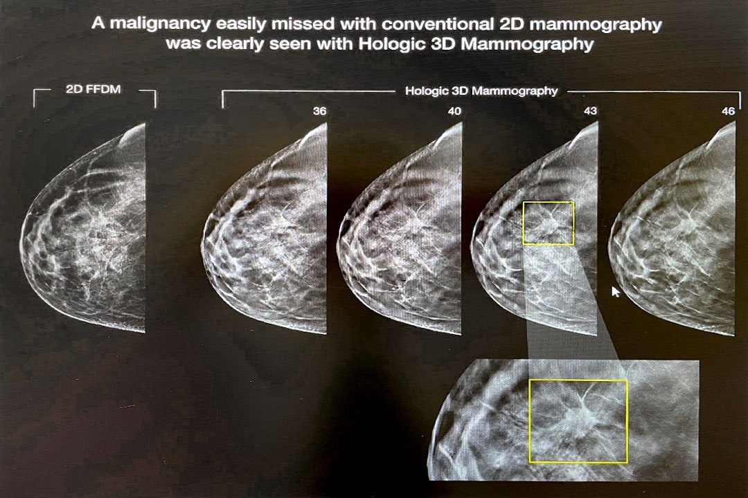
 Our clinics have been performing 3D mammography, also known as digital breast tomosynthesis, since 2018. A 3D mammogram combines multiple breast x-rays to create a three-dimensional picture of the breast. The 3D images show each layer of the breast tissue, one millimeter slice at a time. The images are similar to photographing each page in a book, rather than the front and sides.
Combining 3D mammograms with the standard 2D mammograms reduces the need for additional imaging, increases the number of cancers detected during screening, and reduces the number of false-positives (which means the mammogram looks abnormal, even though there is no cancer in the breast).
What is a breast biopsy?
A breast biopsy is requested when an abnormality is seen on a mammogram or on a breast ultrasound that looks suspicious for cancer. Approximately 10% of women return for additional imaging following their screening mammogram. Eight to 10% have a biopsy and 80% of those biopsy results come back benign (non-cancerous). Source: American Cancer Society
1 of 8 women will develop breast cancer.
The American Cancer Society reports that one out of every eight women (13%) will develop breast cancer in her lifetime. One of every 39 (3%) will die from breast cancer. You may not have a known family history, but cancer can potentially affect anyone. Approximately 75% of those diagnosed with breast cancer have no family history.